

MRI Study Reveals Brain Differences in Patients with Migraine, Offering Hope for Better Diagnostics and Treatment
Posted on April 18 2024,
Stay in the Loop on the Latest in Migraine:


A recent study published in the journal Neurology has found interesting observations on the brain structure of people with migraine. The study compared MRI scans of 296 migraine patients to 155 healthy controls, analyzing cortical surface area, thickness, and volume.
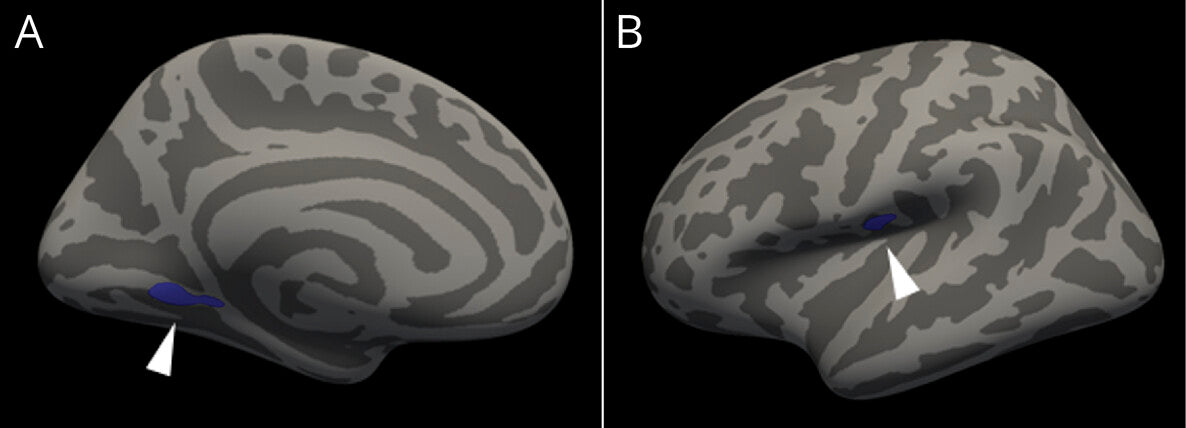
The researchers found that migraine patients, especially those with chronic migraine, had reduced cortical surface area in the left anterior insula, a region involved in pain processing. They also discovered increased surface area in the right caudal anterior cingulate cortex (ACC) in chronic migraine, which correlated with monthly headache frequency. Furthermore, migraine without aura was associated with greater right caudal ACC surface area compared to controls.
No significant differences in cortical thickness or volume were found between migraine and control groups.
The study's findings have important implications for people with migraine. The distinct brain signatures observed in different migraine subtypes suggest that personalized diagnostics and therapies may be possible in the future.
The association between medication overuse/adaptation headache and brain structure changes also highlights the need for effective preventive treatments.
Finally, this is also further evidence that migraine is not “just in your head” as some therapy people we recently talked about like to claim. It is not fear-based, but real, anatomical, and functional changes occurring in the nervous system, including the brain.

Sat, May 30, 26
Candesartan for Migraine Prevention: What New Research Shows
New 2026 research shows candesartan, a low-cost blood pressure pill, can prevent migraines. Here is what the evidence says about how ARBs work, dosing, safety, and who should consider them.
Read MoreFri, May 08, 26
Does the Side of Your Migraine Actually Matter?
If you've been told your migraine attacks are damaging your brain or that the side they're on predicts anxiety or PTSD, the evidence doesn't support it.
Read MoreThu, Mar 05, 26
Migraine, Menopause, and Hormonal Health
Migraine affects women three times more than men, and menopause does not always bring relief. Nearly half of women continue having migraine attacks after menopause. Learn what the research says...
Read More